Then you can perform first measurements to support funding for further developement.
How can an inexpensive NeutronOptics Camera compete, and what more do you need ?
A large variety of CMOS detectors can be used, depending on application.
All cameras are supplied with software & high efficiency x-ray or neutron scintillators.
Try our ImageJ Barrel Distortion Correction macro & example image stack.
Notes on the kind of computer you can use including a Raspberry-Pi (Jesus Mendoza).
We use readily available components that can easily be replaced, even by the end-user.
NEW ! Try the fitsCap imageJ camera macro tool
fitsCap works for all NeutronOptics blue ToupTek and red ZWO detectors (and others), setting capture options, and opening and displaying images directly in imageJ.- Control and plot temperature, with unlimited binning in "sum" or "average" mode
- Execute a command script after each image e.g. to change sample orientation
- Automatically sum or average a series of images; stop when intensity is sufficient
- Automatically subtract calculated diffuse background, or a background file
But click on the links below for instructions for particular NeutronOptics cameras.
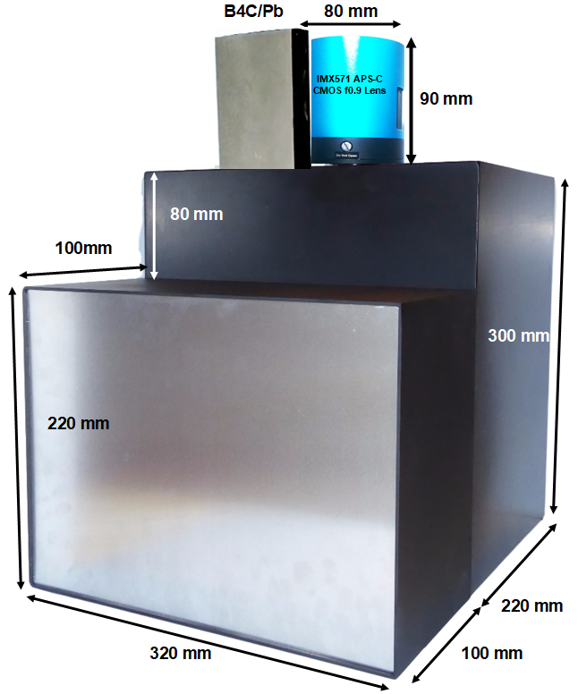
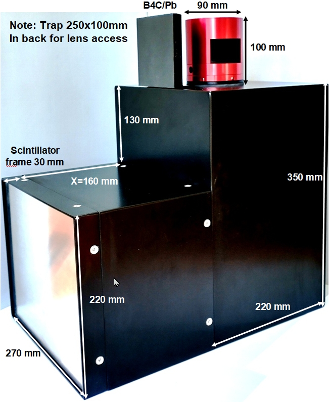
New 300x200mm Efficient High Resolution Camera
Our new standard camera is faster and has higher resolution than our old 250x200mm camera, with a larger 300x200mm FOV in a more compact case. This is achieved by using a larger APS-C CMOS detector with a brighter shorter focal length lens (f/0.95 at 25mm). It maintains the versatility of in-situ interchangeable x-ray and neutron scintillators, while keeping the price below €10K for a large camera complete with scintillator, software and everything needed for immediate operation.Extra lead or concrete gamma shielding is recommended around the detector.
Upgrade old FS60/VS60 CCD cameras to APS-C CMOS
Our old CCD imaging cameras based on the 1" Sony ICX694 12.5x10mm detector with the FS60/VS60 using a fast f/1.4 lens, can be upgraded to our 24x16mm APS-C IMX571 detector with an even faster f/0.95 lens. This is interesting for very low intensity applications, such as very high resolution, fast neutron imaging, Laue backscattered x-ray cameras etc.The x6 speed gain with this upgrade can be summarised as follows.
3D Tomographic Reconstruction
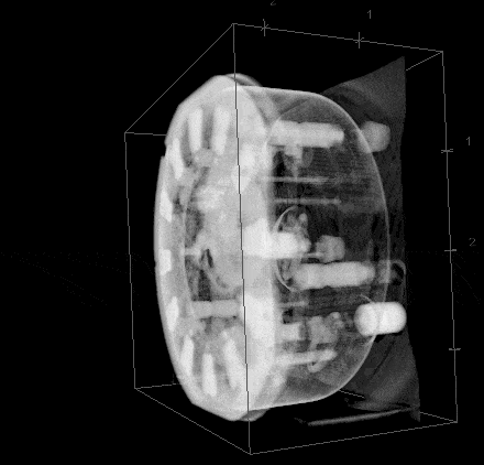 Tomographic Reconstruction requires efficient cameras because many radioagraphs must be collected by rotating the sample at small angular intervals. We recommend the Newport Micro-Controle URS turntables for sample alignment and rotation, and provide simple scripts to synchronise turntable rotation stepping with radiographic imaging. We are grateful to Anders Kaestner at PSI Switzerland for making his tomographic reconstruction programs freely available, and to Burkhard Schillinger at FRM-2 Munich for providing an excellent neutron tomographic data set for a clock.
Very High Resolution Neutron and X-ray Cameras
Very high resolution (<25 µm) requires thin scintillators, which are not very bright, so the camera itself must be very efficient. For small fields-of-View (FOV) of 20-30 mm this is best achieved with our 1:1 Tandem Macro camera, but otherwise we must maximise the ratio of the area of the sensor to the area of the FOV, using bright close-focus lenses; the aperture of a normal macro lens is typically f/2.8, which is only 25% as bright as f/1.0. Our cooled 24x16mm Sony IMX571 sensor with an f/0.95 APS-C lens is ideal for a high resolution camera with an optical resolution of <25 µm over 125x100 mm.

![]()
A B4C/Pb bolt-on detector shield is recommended.
We can provide it with either 100 µm RC-TriTec 6LiF/ZnS scintillators, or even higher resolution RC-TriTec 20 µm Gd2O2S2:Tb scintillators. The GadOx scintillators are only ~8% as bright as the 100 µm 6LiF/ZnS scintillators, but can achieve a resolution of 25 µm.
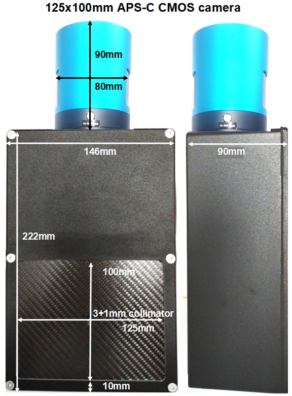
125x100mm Cooled APS-C CMOS Laue Camera
The photo below shows our new 125x100mm CMOS camera, which is the same size, but x6 more efficient than our old 1-CCD Laue camera for the same price. Over 25 years, we have developed various Laue crystal alignment cameras for neutrons and x-rays. They allow rapid crystal alignment, and can also be used for hands-on teaching of crystallography. A finely collimated white beam produces a number of "Bragg spots" from a single crystal, and by measuring the positions of these spots the crystal orientation can be determined. Greater precision is obtained with backscattering, but the intensities are weaker, especially for x-rays because of the scattering "form-factor". A classic bench-top x-ray generator with a spot size of ~1mm and power of 30-50 kV and 30-50 mA is required.

The W backscattered Laue pattern (above left) was obtained in only 30s on a 2kW generator with W cathode operated at ~35keV with this more efficient 1-CMOS camera. The
CLIP refined fit shows the crystal was oriented to 0.25o.
This German industrial customer converted from an Image Plate Laue diffractometer. See the CMOS Laue camera manual
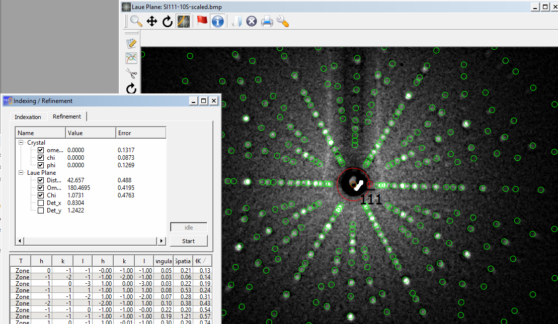
The Si pattern below was collected in only 10s by another user, and oriented and refined with CLIP (Cologne Laue Indexing Program). All spots fit well, and the calculated errors in the crystal orientation are only ~0.1 degrees. (Click the image to enlarge it).
180x120mm Cooled APS-C CMOS Laue Camera
This large 1-CMOS Laue camera is designed to replace our old 2-CCD Laue camera. By using a larger APS-C 24x16mm CMOS detector instead of two 12.5x10mm CCDs, we increase the total detector area (efficiency) by over 50% allowing a slightly larger Field-Of-View (FOV) for similar exposure times (3-5 minutes). The efficiency is further improved by using a modern f/0.95 lens instead of a pair of f/1.4 lenses. The incident beam collimator (1mm/2mm) passes through a mirror and a scintillator in the centre of the camera, and the backscattered diffraction pattern is collected in real time for precise crystal orientation. This new camera with higher efficiency and simpler operation is also less expensive !

![]()
The dimensions can be adjusted to order.
See the CMOS Laue camera manual
High efficiency & High Resolution Macro Cameras
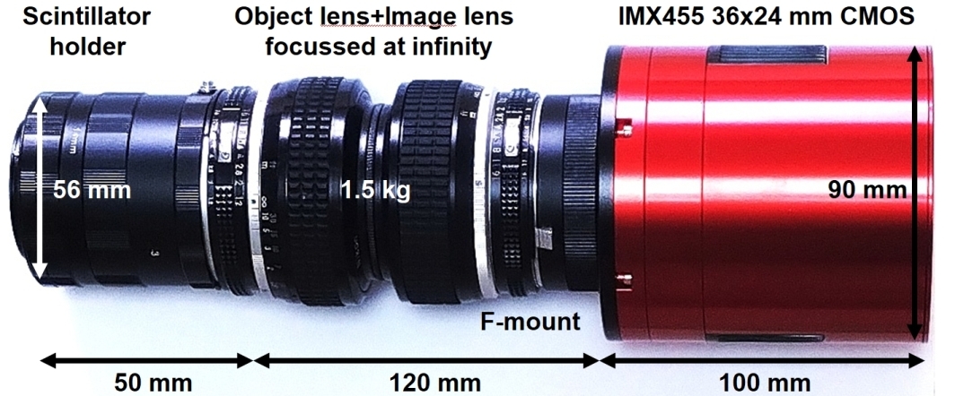
Macro cameras with a small Field-of-View (FOV) are highly efficient, with high resolution, especially our 1:1 Tandem macro camera. It is shown above without the 90-degree mirror, to reduce the distance between the lenses and increase the FOV. It uses a 36x24mm IMX455 CMOS detector with a pair of bright Nikkor f/1.2 full frame lenses to give a relatively large 30x24mm FOV with 3.75 µm pixels. This camera is suited to soft x-ray imaging when used with an ultra-fine GadOx scintillator with a very thin 30 nm aluminium window coating.
A less expensive APS-C version, with a correspondingly smaller FOV, is also available.
High Resolution Tandem 1:1 and 3:1 Macro cameras
 As an improvement to our 1:1 macro camera, we can propose a Tandem Macro option, using a 35mm f/1.2 imaging lens in front of the 100mm lens with a tiny IMX249 CMOS detector (5.86µ pixels). Both lenses are focussed to infinity to give a 3:1 macro image of our 50µ wire grid with <2µ pixels
x2 brighter than with our normal 1:1 macro camera.
We can use other tandem macro imaging lenses of different focal lengths depending on the required magnification and intensity. For example, this pair of Nikon Nikkor 50mm f/1.2 tandem lenses is x8 brighter than our normal macro camera, with the same 1:1 resolution. Maximising intensity is important when thin scintillators are used for the highest resolution. (Williams et al. and our Rodenstock camera).
Tandem macro lenses are claimed by PCO to be competitive in efficiency to fibre-optic bundles bonded directly to the chip, combining high efficiency with high resolution, but the FOV is small (~8mm diameter for our 1:1 Tandem Macro camera) .
The image on the right was obtained on the ILL NeXT D50 beamline from our Twin Nikkor 50mm 1:1 camera using a 10µ thick Gd2O2S:Tb/6LiF PSI/RC-TriTec scintillator with 6% of the light output of their 200µ scintillator. The inner circle shows that at least 25µ resolution was obtained for an exposure of only 3s with a neutron flux estimated to be ~5x10**7 n/cm2/s.
Gd2O2S is also a good x-ray scintillator. (Credits: Alessandro Tengattini & Lukas Helfen, ILL)
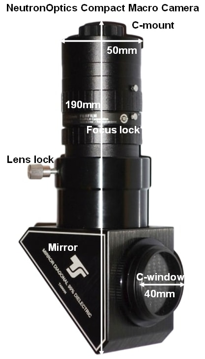
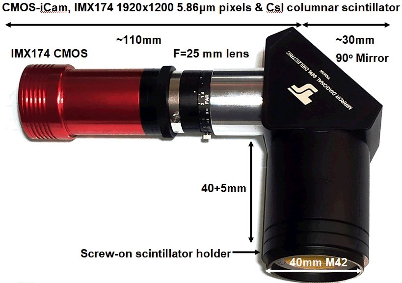
Compact CMOS x-ray or neutron cameras
USB powered CMOS detectors can be used instead of our "slim" CCD for both iCam and larger cameras. The CMOS iCam (right) like the compact iCam has ~25µM resolution over a 40mm FOV.
We can use the same compact CMOS detector in place of our old "slim" CCD in any of our small x-ray or neutron detectors, such as the 100x100mm x-ray camera (left), increasing the resolution to 1920x1200 pixels, or ~85µM. (Real resolution depends on the scintillator).
Fast Neutron Radiography with our Cameras
Fast neutrons (2.45MeV or 14MeV) are very much harder to detect than thermal neutrons because they are not captured by nuclei to provide ionising fission fragments. Scintillation in ZnS can however be achieved with knock-on protons rather than 6Li fission, though long exposures are needed. Fast neutron scintillators use high density polypropylene PP as a source of knock-on protons, and NeutronOptics now offer RC-TriTec PP-ZnS scintillators. Standard sizes are 75x50mm, 125x100mm and 250x200mm (smaller is more efficient).Extra lead or concrete gamma shielding is recommended around the detector.
 Small D-D and D-T neutron generators are a promising development for low-cost neutron radiography, though the fast neutron flux is low. Even so, one of our 250x200mm cameras has already been used with an AdelphiTech 2.45 MeV D-D neutron generator to obtain radiographs with long exposures (~10 min).
The efficiency of the camera can be increased by a factor of x4 by reducing the FOV to 125x100mm, and can be further increased by a factor of x64 by binning pixels 8x8. Since the resolution after binning the smaller FOV will still be 360µm, this is still sufficient with the 2.5mm PP scintillators used for fast neutrons. The smaller camera is shown above. A cheaper compact version is also available.
Other clients from ETH-Zurich, PSI Switzerland and the Hungarian Academy of Science have recently used our smaller TS14 camera for fast neutron radiography and tomography at the 10 MW Hungarian reactor, using an 8 mm thick BC400 transparent plastic scintillator with spatial resolution of around 1.3 mm. It is remarkable that images were obtained through massive 30 cm thick lead shielding and filtering to attenuate gamma background.
An Experts Meeting on Fast Neutron Radiography at FRM2 Munich in October 2019 summarises progress, and includes our paper on An Efficient Camera for Fast Neutrons.
![]()
Test of a miniature x-ray imaging system
X-ray tomographic imaging with a miniature fine focus x-ray generator was recently tested at an international agency in Vienna. Our 125x100mm camera was used with a benchtop MOXTEK fine focus x-ray source and an inexpensive CAWO OG2 fine plastic x-ray scintillator. The palm-sized x-ray source was enclosed in the small shielded grey box (below). Operating at 30KV with a few micro-amps provided sufficient intensity for radiography with background radiation of <2mR/hr (similar to radiation levels in an aircraft cabin). Ease of use and low radiation levels are important for the intended use in training students.
With the higher power available from these miniature sources, with still minimal x-ray shielding, it would be possible to obtain geometrically magnified images of up to x10 by moving the object further from the scintillator screen. This requires a divergent beam and fine focus source to avoid image blurring. (AMETEK also make miniature x-ray sources, but with larger spot size). Other larger fine-focus generators such as the Oxford Instruments Ultrabright are also suitable. With high resolution scintillators in our macro camera, it would be possible to obtain micron-level resolution with high magnification from a divergent micro-focus source.
Big 500x400mm X-ray or Neutron Imaging Camera
We have made 400x300mm and even 500x400mm imaging cameras with a full-frame 36x24mm detector, such as the new Sony full-frame IMX455 CMOS chip, which with 9576x6388 pixels is capable of an optical resolution of better than 100 µm. The 500x400mm camera is a 2x2 scaled up version of our standard 250x200mm imaging camera, which itself is 2x2 times bigger than our compact 125x100mm camera (photos below). Remember that the efficiency of the camerta is proportional to the ratio of the sensor size to the FOV, so bigger cameras are less sensitive for the same sensor (and the scintillator costs much more).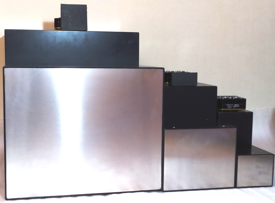
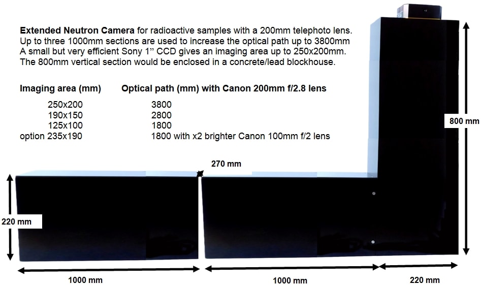
Extended Camera for Radioactive Samples
Radioactive samples require special precautions to avoid damage to the detector. Our solution is to use a telephoto lens to increase the optical path, so that the detector section can be housed in a blockhouse several meters from the active sample, which can then be close to the scintillator to obtain the best resolution. This extended L-shaped camera is a stretched version of our 250x200mm "fast" camera. Relatively inexpensive 200mm or 100mm Canon or Nikon lenses are used, though these lenses with f/2.0 or f/2.8 are not as bright as our usual f/1.2 or f/1.4 lenses; an f/1.4 lens is x4 as efficient as an f/2.8 lens, but this camera is still fast enough for tomography on a low flux Triga reactor.Our "extended" camera has up to three 1000mm long front segments with an 800mm high vertical section holding the detector, lens and mirror to give a maximum optical path of 3800mm, and a minimum of 800mm. Standard Canon or Nikon lens mounts are used, so it is easy to change the lens as well as the optical path.
 (Click on the photos to enlarge).
Controlling CMOS imaging cameras via Ethernet & WiFi
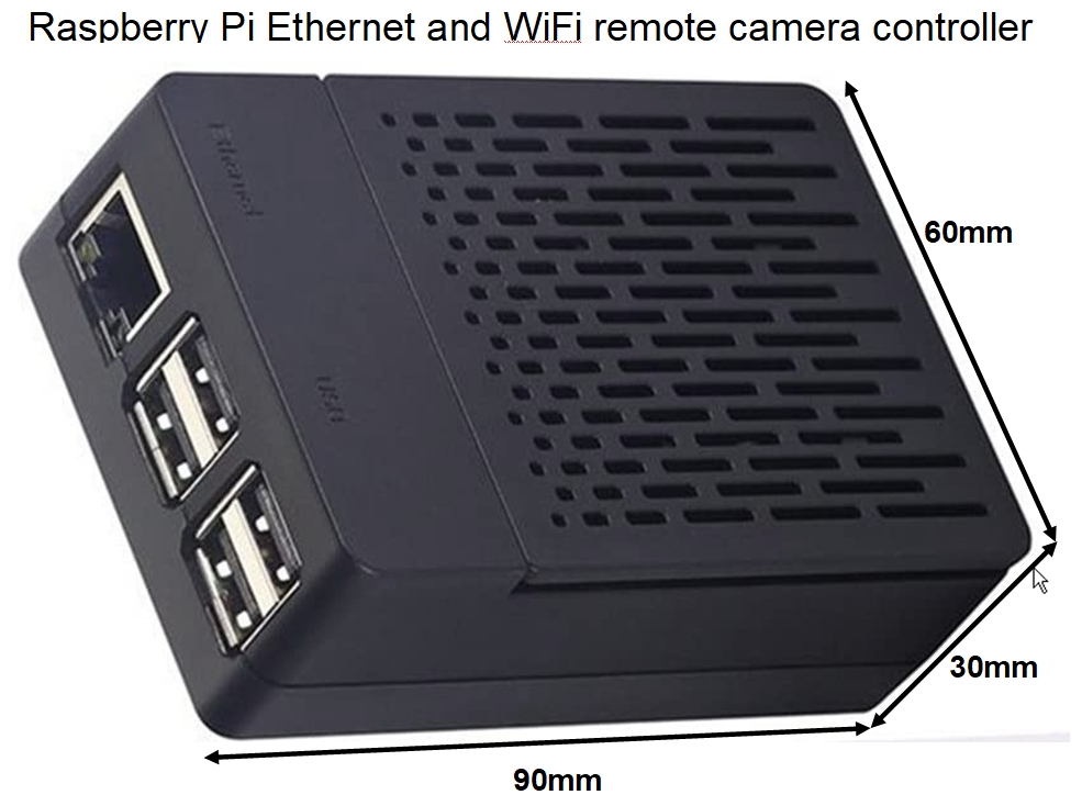 Our small cameras can be supplied with slim GigE Power-Over-Ethernet CMOS detectors. Otherwise
we supply 10m amplified USB cables with all USB cameras, and they can be chained up to 30m. For our larger imaging cameras, we also offer a Raspberry Pi nano-computer as an interface with Atik Air software to allow remote control over Ethernet or WiFi.
It uses a 1.2 GHz 64-bit CPU with 1 GB RAM and is designed as an inexpensive (<€100) general purpose computer with Ethernet, WiFi, HDMI video and external USB storage, but we use it only as an interface.
Our small cameras can be supplied with slim GigE Power-Over-Ethernet CMOS detectors. Otherwise
we supply 10m amplified USB cables with all USB cameras, and they can be chained up to 30m. For our larger imaging cameras, we also offer a Raspberry Pi nano-computer as an interface with Atik Air software to allow remote control over Ethernet or WiFi.
It uses a 1.2 GHz 64-bit CPU with 1 GB RAM and is designed as an inexpensive (<€100) general purpose computer with Ethernet, WiFi, HDMI video and external USB storage, but we use it only as an interface.
Using our Scintillator Frames with Photographic Film
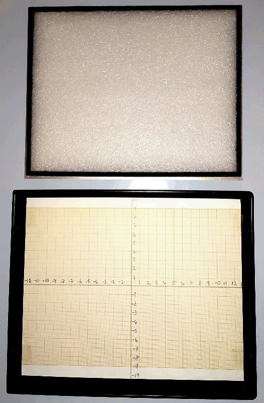 Our camera's scintillator frames can also be used for contact exposures with photographic film. The film is placed on the scintillator in a dark room and covered by our optional camera-frame back. The same scintillator can be used with either a CMOS camera or film.
The bottom image shows the frame with a graph paper focussing scale covering the scintillator. Note the 5mm black B4C rubber seal around the inside of the frame, which normally presses against the camera flange to exclude light. The top image shows the optional film back, which is another 5mm B4C rubber seal on an aluminium plate. This smaller second seal fits inside the seal in the frame to exclude light. This second seal is filled with expanded polystyrene which presses the film gently against the scintillator.
The foam can be covered by a thin B4C layer to prevent thermal neutron backscatter.
(click images to enlarge them)
2D Honeycomb Collimators for Hydrogenous Materials
 Thermal neutrons are strongly scattered by hydrogenous materials, and indeed such organic element contrast is the main advantage of neutron radiography over x-rays. But the scattered neutrons end up in the background, reducing contrast in the detector. Attempts have been made to use 2D honeycomb collimators to reduce such background. Although they are not sufficiently absorbing for fast neutrons, a 50mm long, 5mm wide cell collimator should work for thermal neutrons.
All these cameras use a white neutron beam, and will work on either reactor or spallation neutron sources.
For further details of their application and availability, please contact
Alan.Hewat@NeutronOptics.com.
![]()